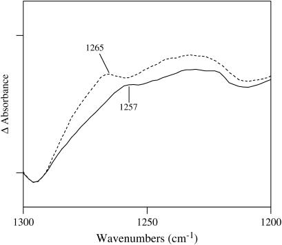
FIGURE 5.
Comparison of the FTIR difference spectra of wild-type and Y34F MnSOD. The tick marks on the ordinate represent 0.02 absorbance units. Note that the intensity at 1265 cm−1 has decreased significantly in Y34F compared with wild-type. These data show that the C-O stretching vibration of the phenolic group of the tyrosine side chain at residue 34 in wild-type makes an infrared contribution at 1265 cm−1. This frequency is characteristic of phenol groups that act as hydrogen bond donors (21).

