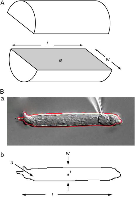
FIGURE 1.
Definition of parameters. (A) OHCs are modeled as cylinders, and the parameters length (l), width (w), and longitudinal section area (a) are defined as shown in the diagram. (B, a) Actual picture of an isolated OHC with its automatically defined boundary delineated in red; (B, b) the automatically defined boundary and the centroid of the cell as measured by DIAS software. The cell width (w) is measured at the cell centroid.

