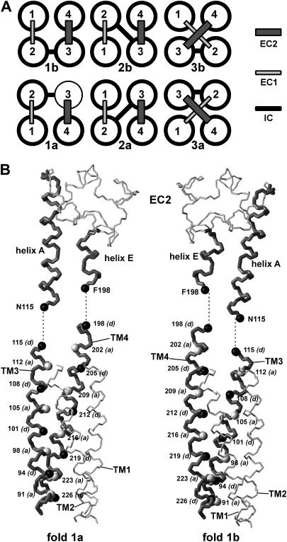
FIGURE 4.
Possible folds of a square four-helix bundle and discrimination between folds 1a and 1b. (A) The six possible folds obtained by permutation of helices. (B) Backbone representation of the EC2 and its connectivity with the modeled transmembrane domain for folds 1a (left) and 1b (right). Transmembrane helices TM3 and TM4 and EC2 helices A and E are drawn in dark gray and thicker bonds. The Cα atoms of the internal coiled coil core residues a and d are, respectively, represented in light gray and black and labeled.

