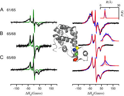
FIGURE 13.
EPR spectra of spin-labeled T4 lysozyme mutants. Shown on the left are the sum of singles spectra (T4L 61 + T4L 65, T4L 65 + T4L 68, T4L 65 + T4L 69; green lines) with the spectra of the double cysteine mutants (T4L 61/65, T4L 65/68, T4L 65/69; black lines) overlaid on an expanded (×5) scale. The spectra of the double cysteine mutants are also shown on the right (black circles) with fits obtained to the tether-in-a-cone model (blue lines) and obtained by the convolution method (red lines) overlaid. The distance distributions obtained using both methods are plotted in the insets at the top right. All spectra were obtained as described in the text with the spin-labeled T4L mutants in desalting buffer plus 70% (w/w) glycerol at −30°C. Analysis of the spectra of the four single cysteine mutants gave the following parameters (fits not shown): T4L 61, gxx = 2.00814, gyy = 2.00606, gzz = 2.00230, Axx = 6.34 Gauss, Ayy = 5.45 Gauss, Azz = 36.29 Gauss, Lorentzian linewidth Γ = 2.28 Gauss; T4L 65, gxx = 2.00821, gyy = 2.00603, gzz = 2.00226, Axx = 6.43 Gauss, Ayy = 5.54 Gauss, Azz = 36.07 Gauss, Γ = 2.31 Gauss; T4L 68, gxx = 2.00817, gyy = 2.00606, gzz = 2.00228, Axx = 6.51 Gauss, Ayy = 5.27 Gauss, Azz = 36.05 Gauss, Γ = 2.31 Gauss; T4L 69, gxx = 2.00815, gyy = 2.00602, gzz = 2.00233, Axx = 6.48 Gauss, Ayy = 5.35 Gauss, Azz = 36.01 Gauss, Γ = 2.30 Gauss. The parameters obtained by fitting the three spectra of the double cysteine mutants to the tether-in-a-cone model are given in Table 2. Fits were obtained with the following parameters fixed: q = 7.08 Å, β = 78.9°, and γ = 330.5°. Values of p were fixed as given in Table 2. The parameters obtained by fitting the spectra of the double cysteine mutants using the convolution method were: T4L 61/65, R0 = 7.51 Å and σR = 0.61 Å; T4L 65/68, R0 = 10.61 Å and σR = 5.07 Å; T4L 65/69, Amplitude1 = 1.00, (R0)1 = 7.51 Å, (σR)1 = 0.61 Å, Amplitude2 = 0.82, (R0)2 = 9.90 Å, and (σR)2 = 5.57 Å. The inset at the center of the figure is a ribbon diagram representing the structure of T4L (PDB, 3LZM (40)) with the α-carbons of residues 61 (red), 65 (green), 68 (blue), and 69 (yellow) shown as colored spheres.

