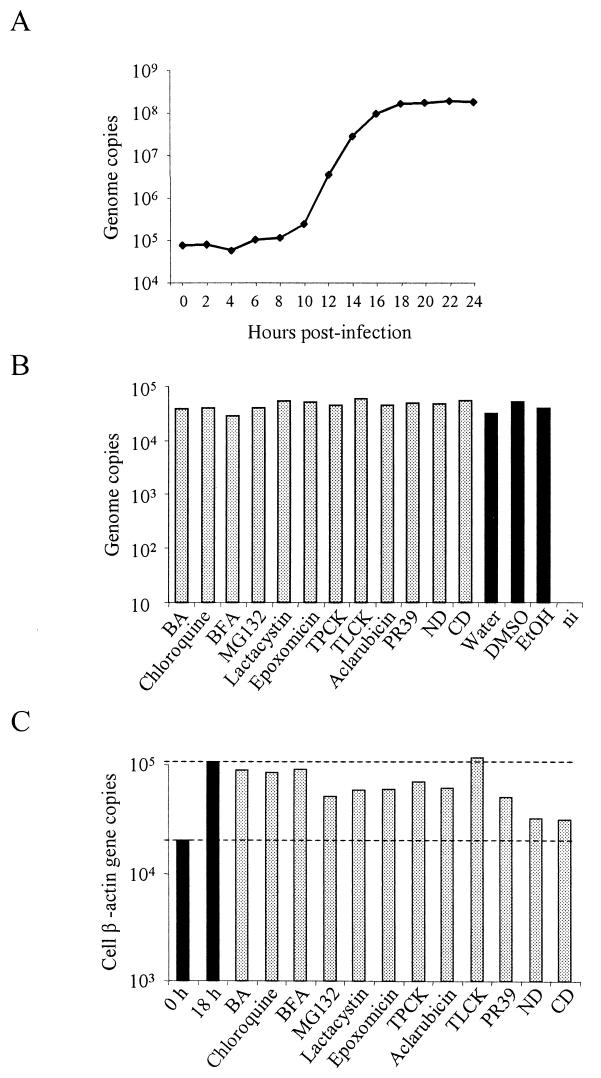
FIG.1.
(A) MVM DNA replication kinetics. A9 cells were infected with MVMp at an MOI of 10 for 1 h at 4°C, followed by washing to remove unbound virus, and were further incubated at 37°C. At 2-h intervals, total DNA was extracted and quantified as described in Materials and Methods. (B) Viral binding and internalization assay. A9 cells were infected in the presence of the highest doses of the different drugs. Cells were then washed and incubated in the presence of the drugs at 37°C for 2 h to allow internalization. Subsequently, cells were collected and were further incubated in a trypsin-EDTA solution to remove viral particles that have not internalized. The cells were pelleted, and after final washings, total DNA was extracted and MVM DNA was quantified by real-time PCR. DMSO, dimethyl sulfoxide. (C) Cell DNA synthesis assay. A9 cells were treated with the highest doses of the different drugs and were incubated at 37°C. At 0 and 18 h of incubation, cellular DNA was measured by quantifying the copy numbers of the mouse β-actin gene (GenBank accession no. M12481) by real-time PCR. Values represent the mean of three (two for panels B and C) independent experiments. ni, noninfected.

