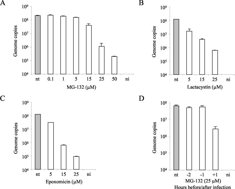
FIG. 5.
Sensitivity of MVMp to proteasome inhibitors. (A to C) Cells were infected with MVMp for 1 h at 37°C at an MOI of 10 in the presence of increasing doses of MG132, lactacystin, or epoxomicin. Subsequently, cells were washed to remove unbound virus particles and were incubated at 37°C for an additional 1 h (3 h for MG132) in the presence of the inhibitors. Cells were washed and were further incubated at 37°C. (D) A9 cells were treated for 2 h with MG132 (25 μM), 2 and 1 h before and 1 h after virus exposure. Cells were washed to remove the drug and were further incubated at 37°C. The amount of viral DNA was quantified 18 h postinfection. Values represent the mean of three independent experiments. nt, nontreated; ni, noninfected.

