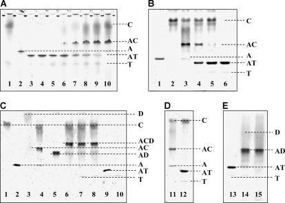
FIGURE 6.
Effect of tβ4 on cofilin-actin and cofilin-actin-DNase I complexes. (Panel A) A representative 10% Native PAGE gel shows the effects of progressive addition of cofilin (C) to the preformed actin-tβ4 complex (AT). (Lane 1) A mixture of 14 μM C and 10 μM tβ4 (T); (Lane 2) 2 μM actin alone (A); (Lanes 3–5) a mixture of 2 μM A and 4, 6, and 10 μM T, respectively; (Lanes 6–9) titration of the preformed AT complex (2 μM A and 10 μM T) with 2, 6, 10, and 14 μM C, respectively; and (Lane 10) a mixture of 2 μM A and 14 μM C. (Panel B) A representative 10% Native PAGE gel shows the reverse order of ABP addition, i.e., progressive addition of T to the preformed AC complex. (Lane 1) A alone (3 μM); (Lane 2) C alone (9 μM); (Lane 3) a mixture of 3 μM A and 9 μM C; (Lanes 4 and 5) addition of 30 and 45 μM T, respectively, to the preformed AC complex (3 μM A and 9 μM C); and (Lane 6) a mixture of 3 μM A and 45 μM T. (Panel C) A representative 10% Native PAGE gel shows C (12 μM), A (3 μM), DNase I (D, 5 μM), and T (45 μM) alone (lanes 1, 2, 3, and 10, respectively). The binary complexes of A with C, D, and T are shown (lanes 4, 5, and 9, respectively). (Lane 6) The ternary complex formed by A, C, and D (A/C/D = 1:1.6:4) in the absence of T. (Lane 7) The addition of a 15-fold molar excess T to the preformed ternary ACD complex (shown in lane 6). (Lane 8) The addition of D to preformed AT (1:15) complex in the presence of a fourfold unbound C (this sample before addition of D is shown in lane 12, Gel D). (Panel D) A representative 10% Native PAGE gel shows the AC (1:4) complex (lane 11), and the addition of a 15-fold molar excess T to it (lane 12). (Panel E) This 10% Native PAGE gel shows the AT (1:15) complex (lane 13); the addition of a 1.6-fold molar excess D to it (lane 14); and the control AD (1:1.6) binary complex (lane 15). Gels were run simultaneously.

