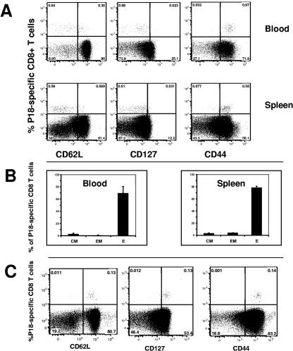
FIG. 5.
Phenotype of HIV-1-specific CD8 T cells elicited by immunization with rM. smegmatis. Mice were immunized with 108 CFU rM. smegmatis expressing gp120 (integrative). (A) Flow cytometric analysis of week 1 PBMC and splenocytes from immunized mice revealed expression of CD44 on the surface of all rM. smegmatis-elicited tetramer-positive cells. CD62L and CD127 were expressed on a subset of the tetramer-positive cells. (B) The proportions of effector (P18-tetramer positive, CD127−, and CD62Llo), effector memory (P18-tetramer positive, CD127+, and CD62Llo), and central memory (P18-tetramer positive, CD127+, and CD62Lhi) cells in the blood and spleen of mice immunized with rM. smegmatis are shown. Effector, effector memory, and central memory cells are denoted E, EM, and CM, respectively. The mean (± SEM) percent E, EM, or CM for each group of mice (n = 4 per group) is shown. (C) Peripheral blood HIV-1-specific CD8+ T cells from mice 1 year after immunization with rM. smegmatis expressing gp120 were predominantly central memory cells. PBMC were pooled from 4 mice that were inoculated twice (10 weeks apart) with 108 CFU bacilli.

