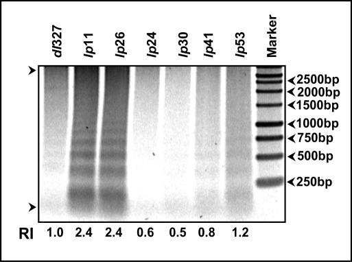
FIG. 6.
Cell death activity. The intracellular low-MW DNA was extracted at 36 h postinfection and analyzed by agarose gel (1.5%) electrophoresis as a measure of cell death activity. The intensity of the DNA fragments in each lane was analyzed with a phosphorimager. RI, relative intensities of the DNA fragments (based on dl327) in the areas indicated between the two arrows.

