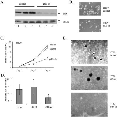
FIG. 6.
pRB depletion inhibits soft agar proliferation of human tumor cells with an activated Ras pathway. (A) Western blot illustrating the decrease in pRB protein levels seen in clones of HT29 cells expressing and pRB-sh (lanes 4 to 6) compared to a MSCV vector (lane 1) or a p16-sh (lanes 2 and 3), using total p44/42 as a loading control. (B) These panels show representative microscopy images of HT29 cells stably expressing a control vector or pRB-sh vector 4 days after plating at 4 × 10⁵ cells/10-cm dish. (C) Clones of HT29 cells transfected with MSCV (vector), p16-sh, or pRB-sh were assayed for proliferation. The empty retroviral vector and a p16-sh expressing vector were used as controls in these experiments since HT29 cells exhibit undetectable levels of expression of p16 due to methylation of its promoter (76). The number of cells in each subline was analyzed at day 2 and day 4 following plating as indicated in the figure. The results reflect the average of two independent experiments performed in duplicate. (D) Analysis of the colony diameter of HT29 foci in cells containing a control vector (one clone), a p16-sh-expressing vector (two clones), or a pRB-sh-expressing vector (two clones). Equal numbers of cells were plated in soft agar for 18 to 20 days after which colonies were photographed. The results presented in the figure are derived from two independent experiments in which the diameter of 50 separate colonies was measured and expressed as an arbitrary size (vector, 20.2 ± −0.5; p16-sh, 23.7 ± −10; pRB-sh, 8.75 ± −0.5). (E) Representative images of HT29 colonies in soft agarose from cells containing a control plasmid, a p16-sh-expressing plasmid, or a pRB-sh-expressing plasmid.

