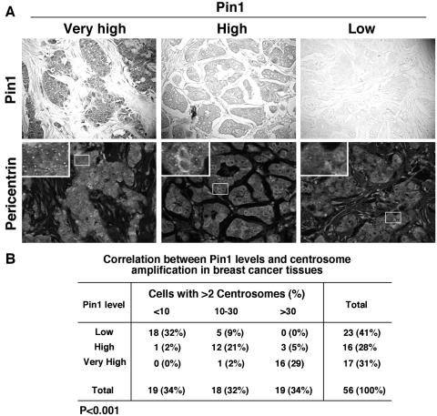
FIG. 1.
Pin1 levels correlate with centrosome amplification in human breast cancer tissues. (A) Breast cancer tissue sections were immunostained using anti-Pin1 antibodies and visualized by DAB staining (upper panels), and different sections of the same tissues were immunostained with antipericentrin antibodies and visualized by Alexa488-conjugated secondary antibody along with propidium iodide to stain nuclei (lower panels). The ratio of centrosomes to nuclei was scored in each sample of >200 nuclei under a fluorescence microscope. The far left panels show a representative very high Pin1 staining, with >30% of cancer cells containing more than two centrosomes, the middle panels show a representative high Pin1 staining with 10 to 30% of cancer cells containing more than two centrosomes, and the right panels show a representative low Pin1 staining with <10% of cancer cells containing more than two centrosomes. (B) The levels of Pin1 expression and the percentage of cancer cells containing more than two centrosomes were determined in 56 surgical specimens of breast cancer as shown in panel A, and their correlation was analyzed by a Spearman rank correlation test (P < 0.001).

