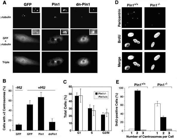
FIG. 3.
Pin1 inhibition or ablation suppresses centrosome duplication in CHO cells and mouse embryonic fibroblasts. (A) and (B) Inhibition of Pin1 suppresses centrosome amplification in S-arrested CHO cells. CHO cells were transfected with plasmids encoding GFP, GFP-Pin1, or GFP-dnPin1 and then treated with hydroxyurea (HU) for 40 h to trigger abnormal centrosome amplification. (A) Centrosome numbers were analyzed quantitatively by immunofluorescence analysis with anti-γ-tubulin antibody and 4′,6′-diamidino-2-phenylindole (DAPI). (B) The percentage of cells containing >2 centrosomes was scored in 300 transfected cells. Error bars, standard deviations from three independent experiments. (C) Pin1+/+ and Pin1−/− MEFs have similar cell cycle profiles at early passages. Exponentially growing primary MEFs derived from Pin1+/+ and Pin1−/− mouse embryos at early passages (below four) were subjected to flow cytometry analysis. Error bars, standard deviations from three independent experiments (D) and (E) Pin1 deletion delays centrosome duplication during S phase in primary fibroblasts. (D) Pin1+/+ and Pin1−/− MEFs were labeled with BrdU for 3 h and coimmunostained with anti-BrdU monoclonal and antipericentrin polyclonal antibodies to analyze centrosome numbers in S-phase cells. (E) For each sample, the number of centrosomes was counted in 200 BrdU-positive cells. Error bars, standard deviations from three independent experiments.

