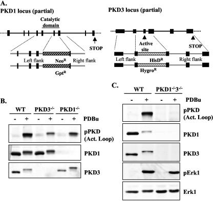
FIG. 1.
Generation of the single and double PKD1 and PKD3 knockout DT40 B cells. (A) Chicken PKD1 and PKD3 genomic loci (partial) were characterized as described in Materials and Methods. (B) Expression and phosphorylation of PKD enzymes in wild-type, PKD1−/−, and PKD3−/− DT40 B cells (either untreated or treated with 25 ng/ml PDBu for 10 min) by Western blotting of whole-cell extracts with the indicated antibodies. (C) Expression and activation of PKD1, PKD3, and Erk1 protein levels in wild-type (WT) and double deficient PKD1/3 DT40 B cells (either untreated or treated with 25 ng/ml PDBu for 10 min) was analyzed by Western blotting of whole-cell extracts with the indicated antibodies. Note: in response to activation, phosphorylated PKD isoforms display reduced immunoreactivity for PKD1 and PKD3 antibodies.

