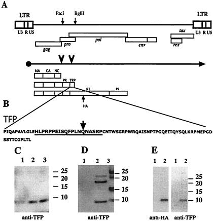
FIG. 1.
TFP is encoded between pro and pol and is a component of the HTLV-1 particle. (A) Major structural feature of the proviral sequences, the primary transcript, and translational products. The unique PacI and BglII sites used in the construction of mutant proviral clones are indicated by small arrows on the genome; the locations of the major genes are shown below the genome. Large arrows on the mRNA transcript mark the frameshift sites. The different precursor proteins are indicated below the mRNA transcript and reflect whether no frameshifts or one or two frameshifts occurred during translation. The mature processing products originating from the precursors are indicated. The position of the HA tag insertion into the pol reading frame is also shown. (B) The sequence of the synthetic peptide within TFP that was used to generate the rabbit antisera. The arrow marks the residue where the frameshift into RT would occur. (C) Immunoblot showing TFP reactive material from different amounts of banded virus from MT2 supernatants. Lanes 1, 2, and 3 contain 1, 2, and 5 μg of total viral protein, respectively. (D) TFP reactive material present in pelleted virus obtained from supernatants of 293 cells transfected with pX1MT (8). A total of 3 × 10⁶ cells were transfected with 8 μg of plasmid DNA by using a calcium phosphate coprecipitation protocol (6). A forty-eight-hour posttransfection virus was pelleted from precleared supernatants for 90 min at 80,000 × g. Lane 1, mock transfection; lane 2, pX1MT; lane 3, marker. (E) Identical bands visible when supernatants from 293 cells transfected with the pCMV-gag-pro-TFPHA construct are probed with either anti-HA or with anti-TFP. Lane 1, mock transfection; lane 2, pCMV-gag-pro-TFPHA. All gels were 4 to 12% NUPAGE gels run in morpholineethanesulfonic acid buffer according to the manufacturer's conditions (Invitrogen, Carlsbad, Calif.). Proteins were transferred to an Immobilon (Amersham) membrane and probed with rabbit anti-TFP or monoclonal rat anti-HA (3F10). Secondary antibodies were coupled with horseradish peroxidase, and reactive material was visualized with Lumiglo (all from Cell Signaling). The positions of the prestained size markers are indicated for each gel.

