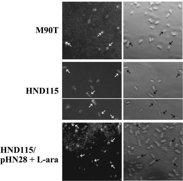
FIG. 4.
Indirect immunofluorescence labeling of IcsA. Panels show IcsA localization using an anti-IcsA antiserum in wild-type strain M90T, in phoN2 deletion strain HND115, and in HND115/pHN28(PBADphoN2) and their corresponding phase-contrast images. In the panels of M90T and HND115/pHN28(PBADphoN2) supplemented with the inducer l-arabinose (L-ara) (PhoN2+), arrows point to representative bacteria exhibiting polar IcsA localization, while in HND115 (apyrase−) arrows point to bacteria presenting IcsA apparently localized over the entire bacterial surface.

