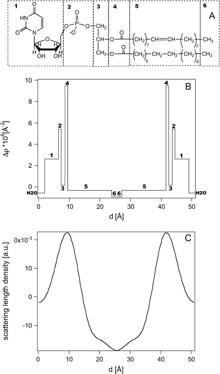
FIGURE 4.
(A) Chemical structure of POPU in which we labeled each submolecualr fragment that contributes to the scattering-length profile. (B) Calculated scattering-length profile evaluated by considering the contributions of different submolecular fragments as separated by sharp interfaces. (C) Experimental scattering-length density profiles at 0% D2O in the direction normal to the membrane plane of the POPU sample.

