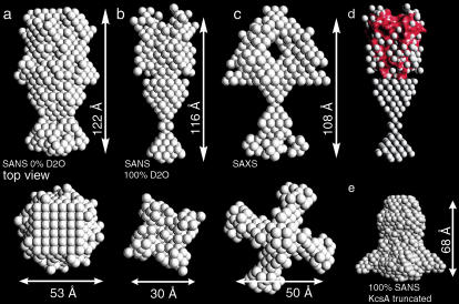
FIGURE 5.
Dummy atom model of full-length KcsA derived from SANS and SAXS intensity data at pH 7.5. Shape models derived from 0% (a) and 100% (b) D2O SANS data are compared the model derived from x-ray scattering (c). (d) A surface representation of the C-terminally truncated KcsA channel (red surface, pdb code 1BL8) aligned with the head domain of the full-length KcsA model derived from the 100% D2O SANS data. The dummy atom radius was reduced to 1.5 Å for clarity. (e) Shape reconstruction from 100% D2O SANS of truncated KcsA at pH 7.5.

