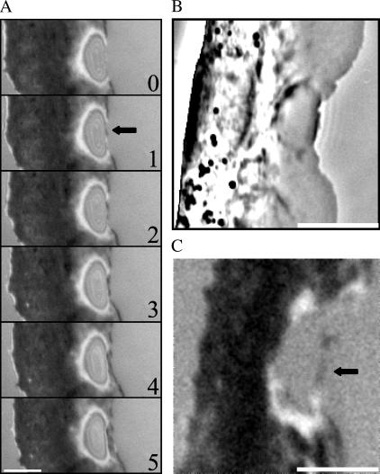
FIGURE 3.
Recovery of the substrate adhesion after the arrest of the leading edge. (A) IRM demonstrates that the adhesion of the leading edge is first reestablished at a small region at the tip of the edge (arrow) and then zips in from both sides, generating a continuous adhesion zone. Simultaneous phase contrast (B) and IRM (C) imaging confirms that adhesion is first reestablished at the very tip of the leading edge. Bar, 5 μm; time in seconds.

