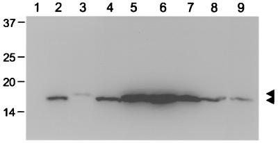
FIG. 2.
Expression and stability of SIVmac239 Vpr protein and derived mutants. COS cells were transfected with 3 μg of plasmid DNA of the empty expression vector (lane 1) or expression constructs for N-terminal Myc-tagged SIVmac239 Vpr (lane 2), HIV-1 NL4-3 Vpr (lane 9), and the SIVmac239 α-helix (lane 3), V21A (lane 4), HxRxG1 (lane 5), HxRxG2 (lane 6), HxRxG1+2 P96T (lane 7), and P96T (lane 8) mutant Vpr proteins. Whole-cell lysates were prepared 48 h after transfection and subsequently separated by electrophoresis. After transfer of proteins to a membrane filter, Vpr proteins were detected by immunoblotting with the Myc monoclonal antibody 9E10 and an enhanced chemiluminescence protocol. The positions of Vpr proteins are indicted by arrowheads, and molecular weight markers are indicated on the left.

