FIG. 5.
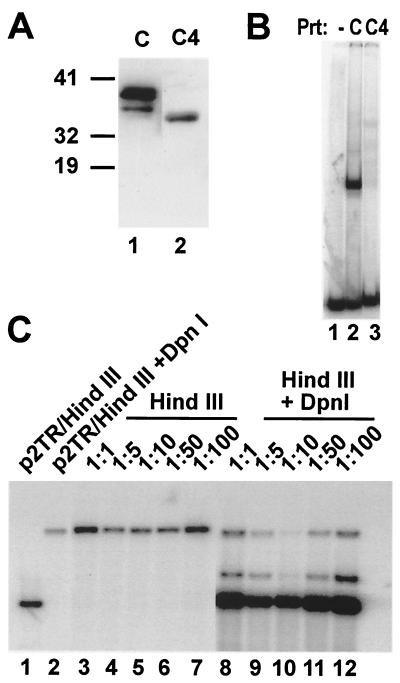
LANA-C4 does not possess dominant-negative activity. (A) Western blot analysis of LANA-C (aa 770 to 1003) and LANA-C4 (aa 785 to 1003) expressed in MVA/T7 vaccinia virus-infected CV1 cells. (B) DNA binding of LANA-C and LANA-C4 to TR5. Equal amounts of recombinant proteins were incubated with radiolabeled TR5 under conditions described in Materials and Methods. Lane 1 contains probe alone, whereas lanes 2 and 3 contain binding reactions with LANA mutants C and C4; C shows strong complex formation, whereas C4 shows extremely weak binding. (C) Titration of increasing amount of pcDNA3.1V5His/LANA-C4 with a constant amount of pcDNA3/ORF73. Short-term replication assays were performed as described for Fig. 1. Then, 100 ng of wt LANA expression construct was cotransfected with a vector expressing LANA-C4 ranging from 100 ng (1:1) to 10 μg (1:100) into 293 cells. Lanes 1 and 2 indicate the positions of DpnI-digested and linearized p2TR. Lanes 3 to 7 contain input DNA, whereas lanes 8 to 12 show newly synthesized DNA. The comparison of input and replicated DNA at each ratio shows that LANA-C4 does not inhibit LANA wt function even when present at a ratio of 100 to 1 (lane 12).

