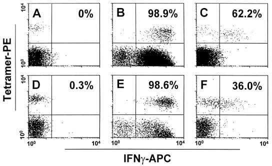
FIG. 2.
IFN-γ expression by p11C tetramer+ CD3+ CD8+ T lymphocytes from peripheral blood and distal-colonic mucosal lymphocytes of a SHIV 89.6P-infected rhesus monkey. Tetramer-binding peripheral blood (A to C) and distal-colonic mucosal (D to F) CD8+ T lymphocytes stain with monoclonal anti-IFN-γ antibody after exposure in vitro to PMA plus ionomycin (B and E) or the Gag epitope peptide p11C (C and F). Percentages of p11C tetramer+ CD3+ CD8+ T lymphocytes positive for IFN-γ are shown in each panel.

