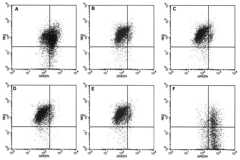
FIG. 3.
Mitochondrial ΔΨm is not altered in reovirus-infected cells. HEK 293 cells were mock-infected (A) or infected with reovirus for 0 h (B), 6 h (C), 12 h (D), or 24 h (E). Control cells were treated with valinomycin (panel F). Cells were incubated with the membrane potential-sensitive dye JC-1 and analyzed using flow cytometry. Red fluorescence indicates mitochondria with intact ΔΨm and green fluorescence indicates loss of ΔΨm. Results are representative of three separate experiments.

