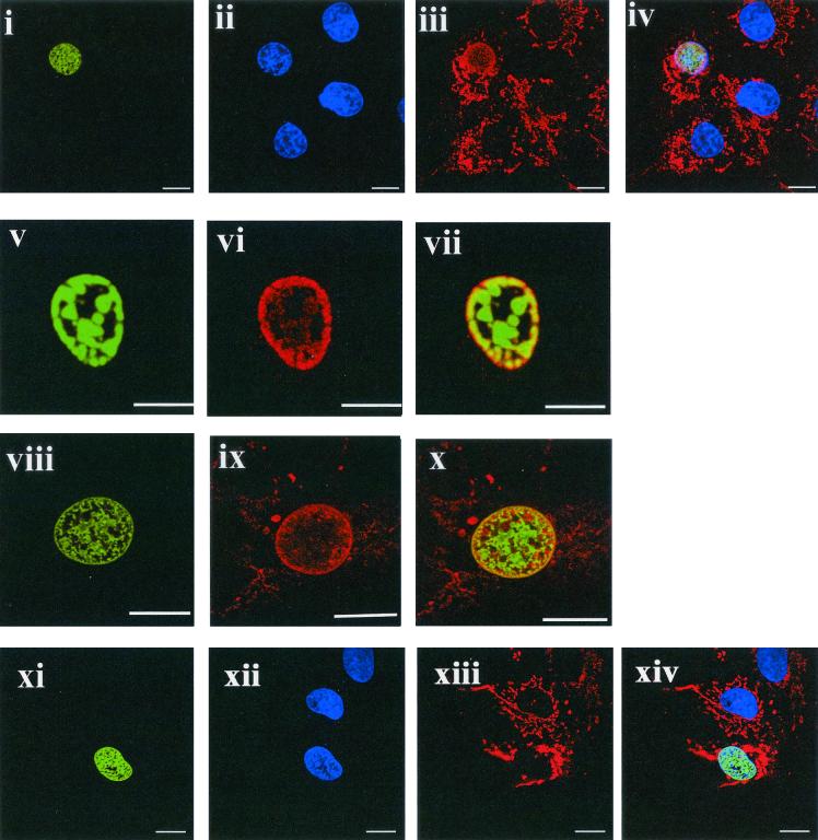
FIG. 4.
The ORF 73 protein accumulates p32 in the nucleus. In all color images, ORF73-GFP or ORF 73 in infected cells stains green, 4; 6-diamidino-2-phenylindole (DAPI) stains blue, and p32 stains red. Images represent a single 0.36-μm focal plane approximately halfway through the cell. Bars, 10 μm. Cells were transfected with p73GFP (i and v), infected with HVS (viii), or transfected with p73ΔNLS1GFP (xi). Each experiment resulted in nuclear localization of ORF73 (or the GFP fusion protein), as shown by DAPI costaining (ii and xii). For infected cells, ORF73 was detected by ORF73 polyclonal antiserum and an anti-rabbit fluorescein thiocyanate conjugate (viii). In all cases, p32 was detected using an p32 antiserum (Santa Cruz) and an anti-goat Texas red conjugate (iii, vi, ix, and xiii). Images iv, vii, x, and xiv are an overlay of the three channels (i to iii), (v to vi), (viii to ix), and (xi to xiii), respectively.

