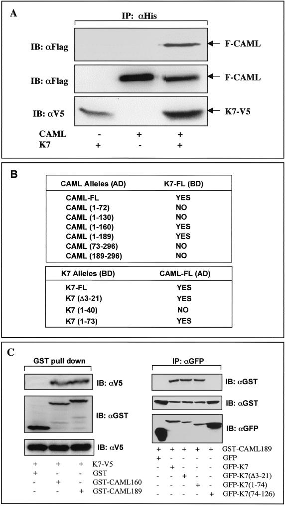
FIG. 4.
Interaction between K7 and CAML. (A) Interaction between K7 and CAML in 293T cells. 293T cells were cotransfected with an expression vector containing V5/His-tagged K7 and/or Flag-tagged CAML. (Top) At 48 h posttransfection, whole-cell lysates were used for immunoprecipitation (IP) with an anti-His antibody, followed by immunoblotting (IB) with an anti-Flag antibody. (Middle and bottom) Whole-cell lysates were also used for immunoblotting with anti-Flag and anti-V5 antibodies to examine the expression of K7 and CAML. Transfection with K7 and CAML expression vector is indicated at the bottom of figure. Arrows indicate V5/His-tagged K7 and Flag-tagged CAML protein. (B) Mapping of regions required for the K7-CAML interaction in the yeast two-hybrid screening. Full-length CAML and its deletion mutants were fused to the Gal4 activation domain (AD) vector, and full-length K7 and its deletion mutants were fused to the Gal4 DNA binding domain (BD) vector. The BD vector containing full-length K7 together with AD vector carrying CAML or its mutants were cotransformed into the AH109 yeast strain. Conversely, the AD vector containing full-length CAML together with the BD vector carrying K7 or its mutants were cotransformed into the AH109 yeast strain. After transformation, AH109 yeast cells were examined for growth on selective Leu-, Trp-, His-, and Ade-deficient plates and for color development on X-α-Gal-containing plate. “Yes” indicates positive results from both assays, and “No” indicates negative results from both assays. None of the transformants showed single positivity in either assay. (C). Mapping of interacting regions of K7 and CAML in 293T cells. (Left) 293T cells were transfected with the GST-CAML mammalian expression vector together with K7 expression vector as indicated at the bottom. At 48 h posttransfection, whole-cell lysates were subjected to precipitation with glutathione-Sepharose beads followed by immunoblotting with an anti-V5 antibody (top). Whole-cell lysates were used for immunoblotting to show the equivalent level of expression of K7 and GST-CAML (middle and bottom). (Right) 293T cells were transfected with the GST-CAML mammalian expression vector together with the GFP-K7 expression vector as indicated at the bottom. At 48 h posttransfection, whole-cell lysates were subjected to immunoprecipitation with an anti-GFP antibody, followed by immunoblotting with an anti-GST antibody (top). Whole-cell lysates were used for immunoblotting to show the equivalent level of expression of GFP-K7 and GFP-K7 mutants (middle and bottom).

