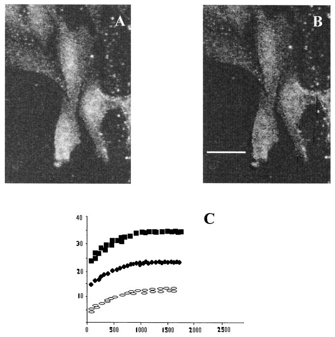
FIG. 5.
FRET measurements for MHC-I and GRP78. Energy transfer between MHC-I and GRP78 can be detected from the increase in donor fluorescence after acceptor photobleaching. (A) Donor (Cy3) image after acceptor photobleaching. (B) E image. (C) E as a function of the fluorescence of donor/acceptor antibody ratios of 1:1 (open ellipse), 1:2 (filled circles), and 1:4 (filled squares). Scale bar, 10 μm.

