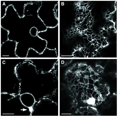
FIG. 7.
Confocal fluorescence micrograph of mock-infected (A and B) or PCV-infected (C and D) epidermal cells of ER-GFP transgenic N. benthamiana. (A) Perinuclear and plasma membranes show a fluorescent halo in a healthy ER-GFP N. benthamiana. (B) Normal reticulate pattern of cortical ER in apical leaf cell of mock-inoculated plants. (C) Perinuclear ER aggregates (arrow) observed in epidermal cells of PCV-infected systemic leaves. (D) Cortical fluorescent bodies and typical ER network in infected leaves. Bar, 10 μm.

