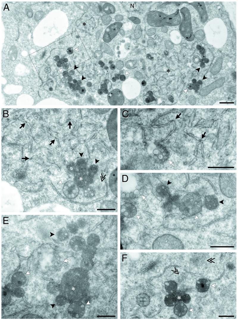
FIG. 8.
Electron microscopy of cytopathological structures in PCV-infected BY-2 protoplasts at 48 hpi. (A) Low-magnification view of the ultrastructural modifications. (B, C, D, E, and F) Higher-magnification views of the different cytopathic structures. Black asterisks correspond to area of broken ER, black arrows point to constrictions on the ER fragments, and white arrows indicate clusters of vesicles. Single arrowheads correspond to MVB; MVB containing disordered membranous vesicles are indicated by black arrowheads, whereas those containing one row of vesicles and surrounded by a single membrane are indicated by white arrowheads. White asterisks correspond to electron-dense material without detectable vesicles. Double arrows indicate clusters of virions. (F) Magnification of the inset square indicated in panel A. N, nucleus. Bars, 1 μm in panel A and 0.5 μm in panels B, C, D, E, and F.

