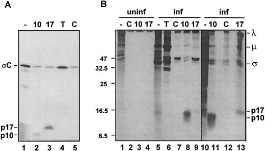
FIG. 4.
ARV S1 mRNA is functionally tricistronic in vitro and in vivo. (A) The full-length ARV-176 S1 cDNA clone was used for the production of capped mRNA by in vitro transcription. The transcripts were translated in rabbit reticulocyte lysates, and the [3H]leucine-labeled translation mixtures were fractionated by SDS-PAGE (15% acrylamide) either without immunoprecipitation (−) or after precipitation with the monospecific anti-p10 (10) or anti-p17 (17) serum, with antiserum raised against total virus structural proteins to detect ςC (T), or with control preimmune serum (C). Radiolabeled polypeptides were detected by fluorography. The locations of the ςC, p17, and p10 gene products are indicated on the left. (B) Uninfected (uninf., lanes 1 to 4) or ARV-infected (inf., lanes 5 to 13) QM5 cells were pulse-labeled with [3H]leucine for 1 h at 16 h postinfection. Cell lysates were fractionated by SDS-PAGE (15% acrylamide) either without immunoprecipitation (−) or after precipitation with the same antisera described for panel A. The locations of molecular size markers are indicated on the left (in kilodaltons), and the locations of the p10, p17, and major λ, μ, and ς virus structural proteins are indicated on the right.

