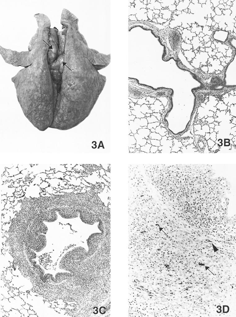
FIG. 3.
(A) Lung from a pig inoculated by the intralymphoid route with PCV2 DNA and necropsied at 21 DPI. The lungs were rubbery, failed to collapse, and were mottled tan-red. Tracheobronchial lymph nodes were markedly enlarged and were tan (arrows). (B) Microscopic section of a normal lung from a control pig (magnification, ×21.25). (C) Microscopic section of the pig lung shown in Fig. 3A. Note the peribronchiolar lymphohistiocytic inflammation and mild necrotizing bronchiolitis (magnification, ×21.25). (D) Immunohistochemical staining of the lung shown in Fig. 3A. Note the PCV2 antigen in macrophages (arrows) and fibroblast-like cells (arrowhead) around airways (magnification, ×54.4).

