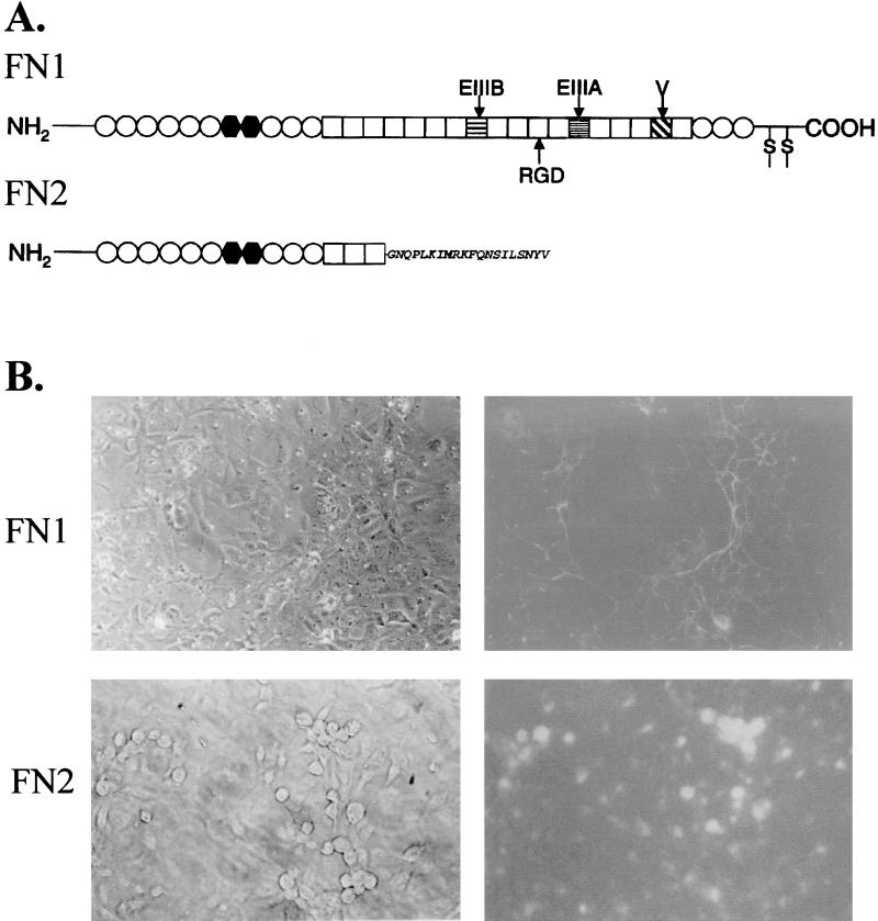
FIG. 1.
(A) Schematic diagram showing the organization of zebrafish FN1 and FN2 (45). The type I (○), II (•), and III (□) repeats along with EIIIA, EIIIB, and the variable region (V) are shown. The amino acid sequence of the FN2 C-terminal tail is indicated. (B) Cellular distribution of recombinant FN1 and FN2, each synthesized as a fusion protein with GFP in CHSE-214 cells. Photomicrographs of the same field examined by light (left) and UV (right) phase-contrast microscopy showing the distribution of the GFP-labeled proteins. Magnification, ×200.

