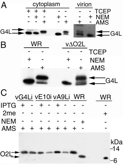
FIG. 1.
Redox status of G4L and O2L. (A) BS-C-1 cells were infected at a multiplicity of 5 with vaccinia virus WR. After 24 h, the cells were TCA precipitated, and the pelleted proteins were solubilized with SDS, treated or not with TCEP, and alkylated with NEM or AMS. Proteins were resolved by electrophoresis in a 10 to 20% polyacrylamide gradient Tricine gel and detected by Western blotting with G4L-specific rabbit antiserum, followed by an anti-rabbit MAb conjugated to peroxidase. Sucrose gradient-purified virions (approximately 108 PFU) were solubilized in 50 mM Tris-HCl (pH 8.0)-0.1% SDS containing NEM or AMS and analyzed as above. The upper arrow points to G4L alkylated with AMS; the lower arrow points to reduced G4L or G4L alkylated with NEM. (B) BS-C-1 cells were infected and the proteins were alkylated as in panel A. Proteins were resolved on a 12% polyacrylamide NuPAGE gel with MOPS buffer and detected as in panel A. The upper arrow points to G4L alkylated with AMS; the lower arrow points to G4L alkylated with NEM. (C) BS-C-1 cells were infected with standard vaccinia virus (WR), vA9Li, vE10Ri, or vG4Li in the presence (+) or absence (−) of IPTG. After 24 h, the cells were TCA precipitated and the solubilized proteins were alkylated with NEM or AMS or reduced with β-mercaptoethanol (2me). Proteins were resolved as in panel B, and O2L was detected with a rabbit antiserum and an anti-rabbit HRPO-conjugated secondary antibody. The upper arrow points to the O2L protein alkylated with AMS; the lower arrow points to reduced O2L or O2L alkylated with NEM. The positions of 14- and 6-kDa protein markers are indicated on the right.

