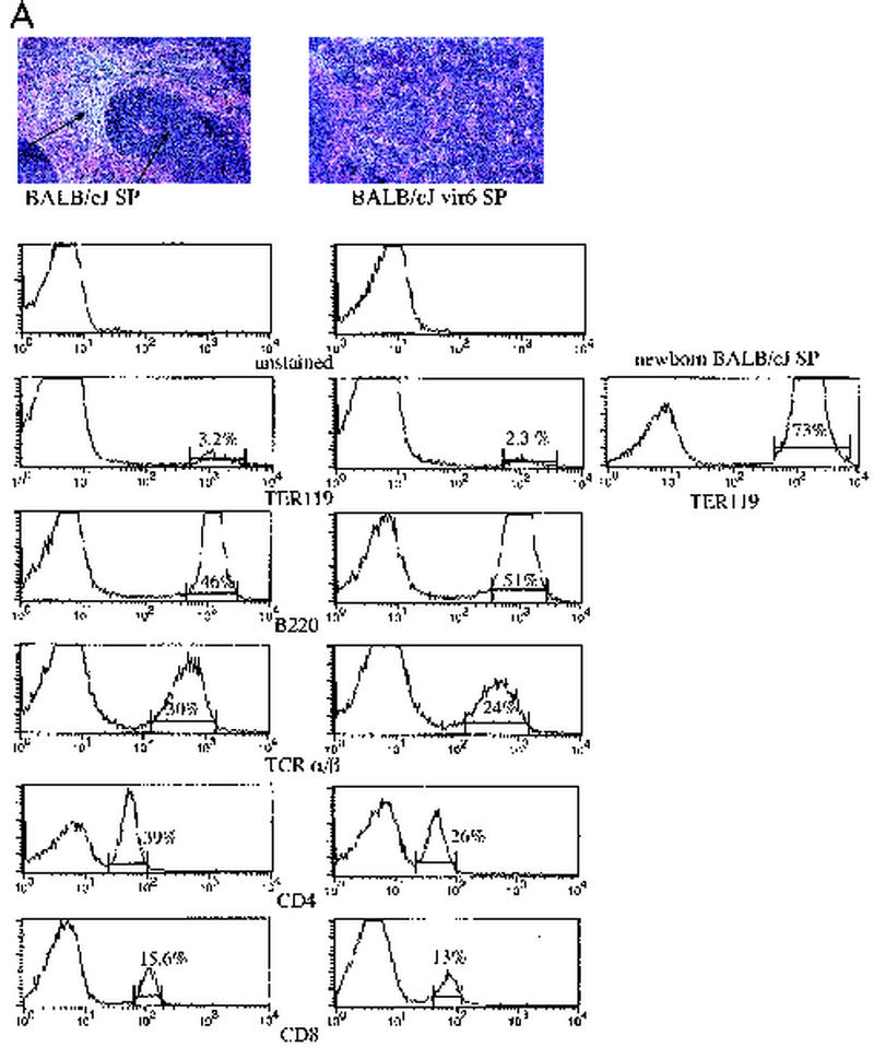
FIG. 1.
Selective proliferation of Sca-1+ cells results in increased extramedullary hematopoiesis and splenomegaly in vir6-infected mice. (A) (Top) Hematoxylin- and eosin-stained sections from spleens of a normal 4.5-month-old BALB/cJ male and an age-matched vir6-infected BALB/cJ male. Arrows, white and red pulps. (Bottom) Flow-cytometric analysis of splenic cells of the same mice. Nucleated spleen cells were stained with anti-Ter119, anti-B220/CD45R, anti-TCR α/β, anti-CD4, and anti-CD8 and analyzed by FACS. Mice infected more than 4 months had enlarged spleens with 160 × 107 to 180 × 107 total cells compared to 6.8 × 10⁷ to 7.6 × 10⁷ total cells in the spleens of uninfected mice. The percentages of cells with surface markers for other cell lineages were also determined, and no differences between infected and control mice were observed (data not shown). Analyzed markers included TCR γ/δ T cells, CD11b/Mac-1 and F4/80 (macrophage specific), NK cells (MAb DX5), and Gr-1/Ly6G (granulocyte specific). To ensure that the osmotic shock used to lyse red blood cells did not affect TER119-positive cells, splenocytes from newborn BALB/cJ mice were included. Data from one of eight experiments are shown. (B) Flow-cytometric analysis of the same splenic cells with anti-Sca-1 MAbs and antibodies against lineage-specific markers. Fifteen infected and 10 uninfected BALB/cJ mice were used. Values are averages ± standard deviations.


