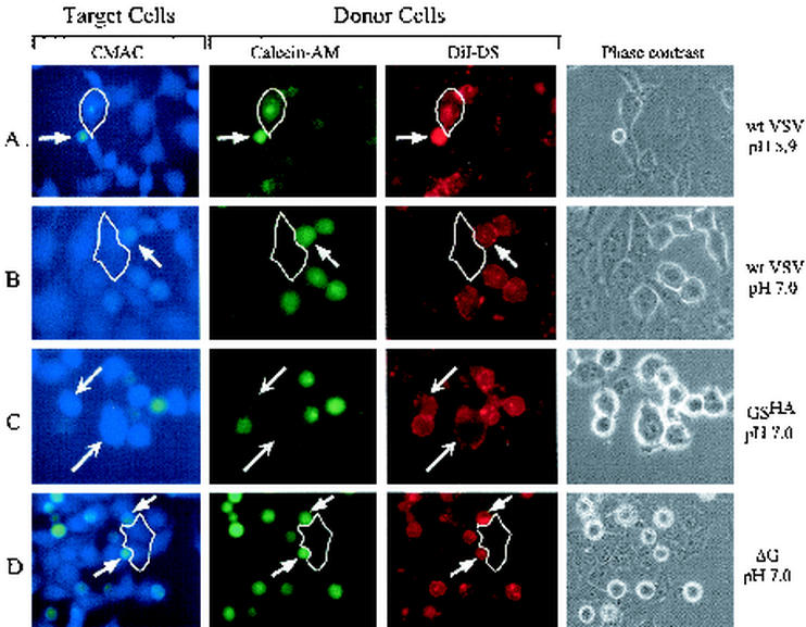
FIG. 6.
Three-color fusion assay. BHK-21 cells were infected with wild-type (wt) VSV (panels A and B), GSHA virus (panel C), or ΔG virus (panel D) at a multiplicity of infection of 10. After 8 h of infection, the cells were labeled with DiI (red) and calcein-AM (green) and then removed from the plates. The cells were then overlaid on BHK-21 cells that had been labeled with CMAC (blue). After the virus-infected donor cells had attached to the plate or settled onto the target cells, the medium was replaced with fusion medium buffered to pH 5.9 or 7.0. After 1 min, the fusion buffer was removed, and the cells were incubated for 20 min in growth medium at 37oC and then placed on ice to prevent any further lipid or content mixing. Phase-contrast and fluorescence images were obtained with filters for 4′,6′-diamidino-2-phenylindole (DAPI) (blue), rhodamine (red), and fluorescein isothiocyanate (FITC) (green). The DAPI filter set did not eliminate bleedthrough of the FITC fluorescence, and therefore the FITC-labeled cells appear greenish-blue in the micrographs. The target cells labeled with CMAC appear as larger, flat blue cells, while the donor cells appear as small, round cells and are labeled both red and green. Relevant target cells are highlighted by a thin white outline in panels A, B, and D. (A) Fusion mediated by wild-type G protein at pH 5.9. The phase-contrast image shows one small donor cell adjacent to two target cells. Transfer of both DiI and calcein-AM from the donor cell to the target cell (blue) can be seen (arrows). (B) A second plate identical to the one shown in panel A but maintained at pH 7.0 shows a lack of transfer of DiI or calcein-AM to the outlined target cell, consistent with the requirement for low pH in initiating VSV G-mediated membrane fusion. (C) Fusion mediated by GSHA at pH 7.0. Several donor cells in contact with target cells are shown. Only the transfer of DiI from the donor to the target cells was seen (arrows). There was no transfer of calcein-AM, showing that complete fusion had not occurred. (D) Multiple donor cells infected with ΔG-VSV which are in contact with an outlined blue target cell are shown, but no transfer of either DiI or calcein-AM occurred.

