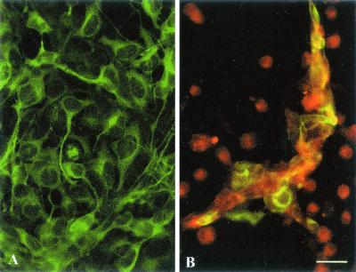
FIG. 2.
Coculture of ScGT1-1 cells and DC. (A) ScGT1-1 cells, immunolabeled with anti-β-tubulin antibodies (green); (B) ScGT1-1 cells (green to yellowish) exposed to DC, ratio 1:1, immunolabeled with anti-MHC class II antibodies (red), for 48 h. Note the extensive loss of GT1-1 cells. Remnants are surrounded by clusters of DC. Scale bar, 25 μm.

