FIG. 3.
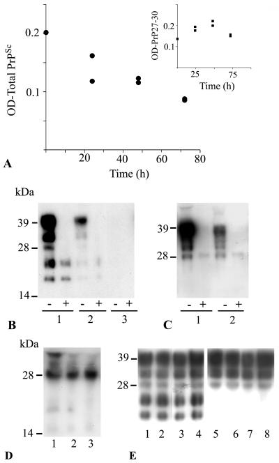
Effect of DC on PrPSc derived from ScGT1-1 cells. (A) Intensity of protease-resistant prion protein in PK-treated (•) lysates at different times after exposure of ScGT1-1 cells to DC, ratio 1:1. The inset shows the intensity of the low-molecular-mass bands (sizes between 19 and 27 kDa) in non-PK-treated lysates, standardized to the intensity of β-tubulin, sampled at different time points after exposure of ScGT1-1 cells to DC; ratio 1:1. (B) Western blot, using Fab D13, of cell culture lysates from ScGT1-1 cells (lane 1); ScGT1-1 cells exposed to DC, ratio 1:1, for 72 h (lane 2); and ScGT1-1 cells exposed to DC, ratio 1:2, for 72 h (lane 3). (C) GT1-1 cells (lane 1) and GT1-1 cells exposed to DC, ratio 1:1, for 72 h (lane 2). Symbols: −, non-PK-treated lysates; +, PK-treated lysates. (D) precipitated proteins from the medium collected from cultures of ScGT1-1 cells (lane 1); ScGT1-1 cells exposed to DC, ratio 1:1, for 72 h (lane 2); or ScGT1-1 cells exposed to DC, ratio 1:2, for 72 h (lane 3). (E) Transwell experiment showing no difference in PrP expression between ScGT1-1 cells unexposed (lanes 1 and 2) and exposed (lanes 3 and 4) to DC, ratio 1:1, for 48 h. Exposure to DC did not affect PrPC expression in uninfected GT1-1 cells (lanes 5 and 6 show unexposed GT1-1 cells, and lanes 7 and 8 show GT1-1 cells exposed to DC).

