FIG. 1.
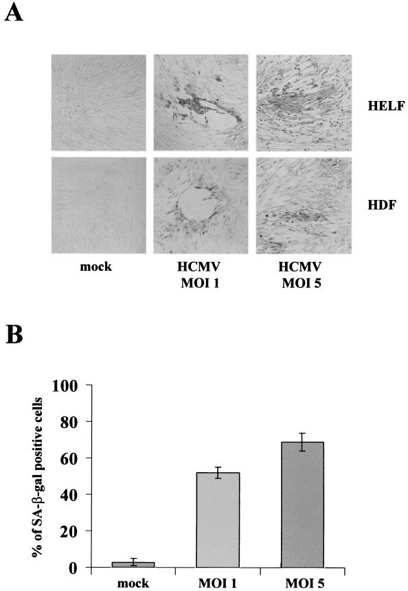
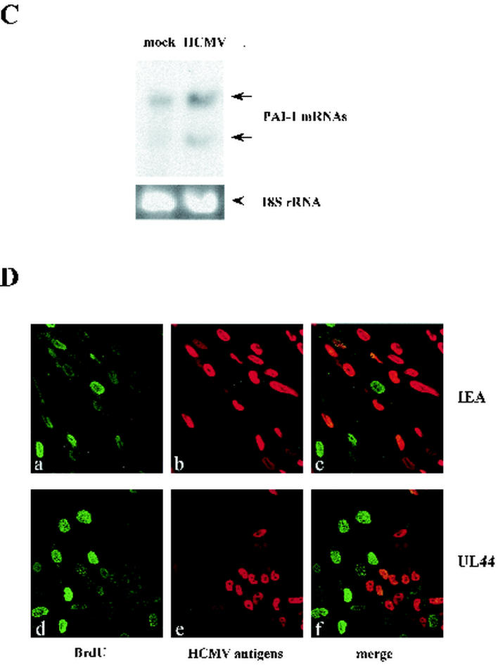
Senescent features of primary fibroblasts infected with HCMV. (A) Histochemical staining of primary fibroblasts infected with HCMV for SA-β-Gal activity. HELF and HDF were serum starved for 72 h, infected with HCMV, stained at 96 h, and photographed. All panels are shown at equal magnification. (B) Percentage of HELF positive for SA-β-Gal at 96 h p.i.. Cells (200) from each cell population were scored; the averages ± standard deviations of data from at least three separate experiments are shown. (C) Expression of PAI-1 in HELF infected with HCMV or mock infected at 24 h p.i.. The two transcripts of PAI-1 (3.2 and 2.2 kb in length) are indicated. (D) Confocal immunofluorescence assays demonstrating that HCMV-infected cells do not incorporate BrdU. After synchronization in the G0 phase by serum starvation, cells were infected with HCMV at an MOI of 1, stimulated with 10% FCS for 24 h, and then pulsed with BrdU for 1 h. Coverslips were processed for immunofluorescence as described in Materials and Methods. Double labeling consisted of a polyclonal anti-BrdU Ab coupled with monoclonal anti-HCMV Abs, either anti-IE antigens (a to c) or anti-UL44 (d to f). BrdU was decorated with a fluorescein isothiocyanate-conjugated secondary Ab (a and d; green), while a Texas-Red-conjugated secondary Ab was used to visualize anti-HCMV Abs (b and e; red). (c and f) Merged images obtained after confocal analysis. Magnification, ×20.


