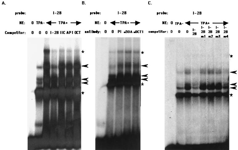
FIG. 6.
EMSA with RE I-2B as a probe. (A) Binding complex of RE I-2B probe and NE. A total of 20 μg of NE from BCBL1 cells induced with TPA or uninduced was incubated with the labeled RE I-2B probe. Each competitor—unlabeled RE I-2B, RE IIC, AP1 (AP1-binding consensus sequence), or Oct (Oct family protein-binding consensus)—shown on the panel was added in a 50-fold molar excess. (B) Supershift analysis with specific antibodies. Next, 20 μg of NE from BCBL1 induced with TPA was incubated with the RE I-2B probe and 2 μg of each of the following antibodies: mouse preimmune serum (PI), α50A (mouse monoclonal anti-RTA antibody), or αOct1 (mouse monoclonal anti-Oct1 antibody). (C) Competition analysis of RE I-2B with its mutants. The labeled RE I-2B probe and each mutant unlabeled probe (in a 50-fold molar excess) were mixed with NE and analyzed. Arrowheads show specifically formed DNA-protein complexes, and the stars indicate nonspecific complexes.

