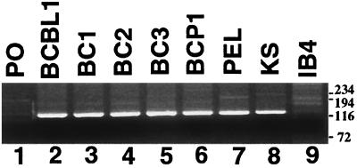
FIG. 5.
RT-PCR of the K12 transcript splice region. RT-PCR with primers flanking the K12 splice sites amplify a 138-bp product after splicing has occurred but would amplify a ∼5-kb fragment in the absence of splicing. Lanes: 1, PO (primers only); 2 to 6, PEL cell lines BCBL-1, BC-1, BC-2, BC-3, and BCP-1, respectively; 7, PEL tumor; 8, KS tumor; 9, IB4 EBV-infected (KSHV-uninfected) lymphoblastoid cell line. Size markers are shown at the right in base pairs.

