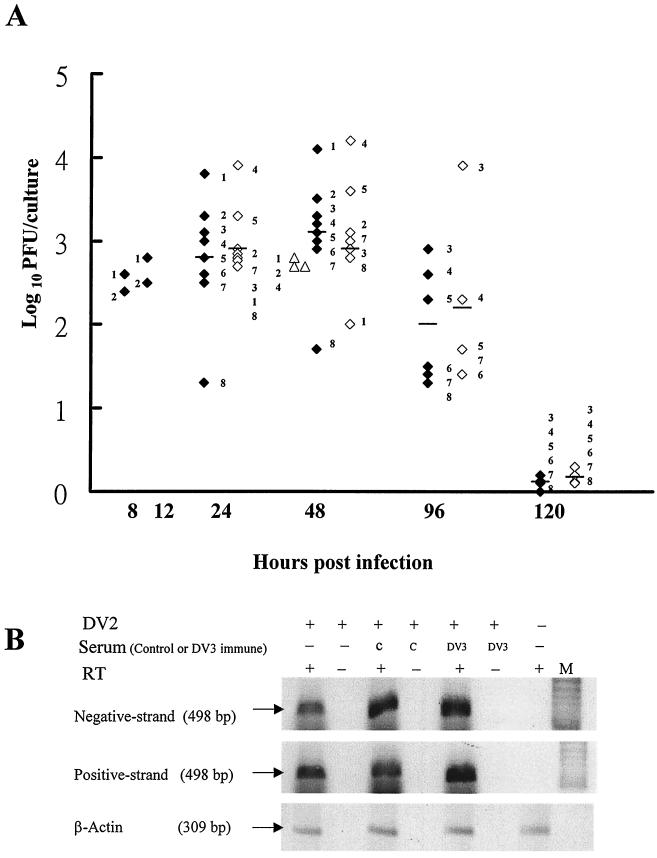
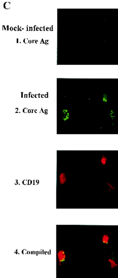
FIG. 1.
Active DV replication in human B cells. (A) B cells were infected with DV2 (solid diamonds) or DV3 (open triangles) and monocytes were infected with DV2 (open diamonds) at an MOI of 10. Culture supernatants were collected at the indicated times to determine virus titers. Numbers adjacent to symbols designate individual donors, and bars represent the mean values for each group. (B) After infection or mock infection of B cells of donor 4 with DV2 at an MOI of 10 in the absence or presence of control or DV3 immune serum, total RNA was isolated from B cells at 24 h p.i. Portions of each sample were hybridized with β2 and D1 primers (top) and the D2 primer (middle) to analyze negative- and positive-strand RNA genomes. Afterward, half of each sample was incubated with (+) or without (−) RT, amplified by PCR, and run in adjacent lanes. β-Actin (bottom) served as an internal control. M, DNA marker. (C) Staining of DV2-infected B cells. After mock treatment (1) or DV2 infection (2 to 4) at an MOI of 10, B cells of donor 6 were collected 48 h later and stained for intracellular viral core protein (1 and 2) and cell surface CD19 molecules (3). Panel 4 is the compiled image of panels 2 and 3.


