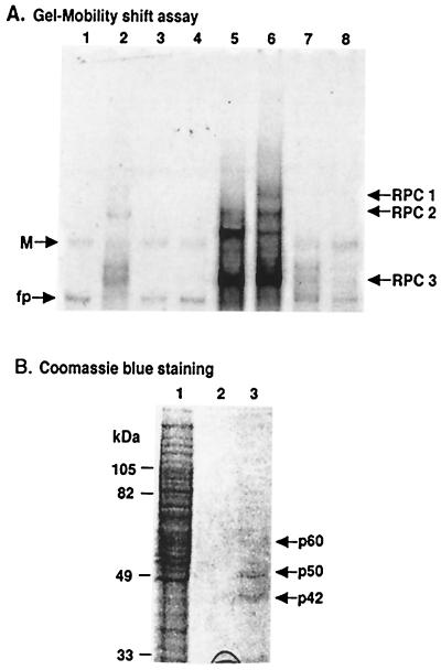
FIG. 1.
Analysis of WNV 3′(−) SL RNA binding proteins in fractions eluted from an agarose-adipic acid hydrazide RNA affinity column. (A) Gel shift assays. Lane 1, free probe; lane 2, final flowthrough fraction from the RNA affinity column; lane 3, first binding buffer wash fraction; lane 4, 0.2 M NaCl wash fraction; lanes 5 and 6, fractions eluted with 1 or 2 M NaCl, respectively; lanes 7 and 8, fractions eluted from a “beads-only” control column with 1 or 2 M NaCl, respectively. For each of the fractions, 1 μl of a total of 100 μl was analyzed on the gel. The positions of the three RPCs are indicated by arrows. M, multimer of the probe; fp, free probe. (B) Coomassie blue staining of the eluted fractions from an agarose-adipic acid hydrazide RNA affinity column. Lane 1, aliquot of sample loaded on the affinity column (10 μl out of 3 ml); lane 2, fraction eluted with 2 M NaCl from a beads-only control column (30 out of 100 μl); lane 3, fraction eluted with 2 M NaCl from the RNA affinity column (30 out of 100 μl). The positions of the eluted proteins are indicated by arrows. The protein markers are shown on the left of the gel.

