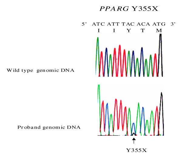
Figure 3.
Genomic DNA sequence electropherograms of heterozygous PPARG mutation. The bottom electropherogram tracing shows both alleles in subject II-5 from kindred 1 (Y355X), compared to sequence from a healthy subject. The position of the mutation is indicated by the arrow. Normal nucleotide and amino acid sequence is shown above the wild type electropherogram tracing.

