
Figure 2.
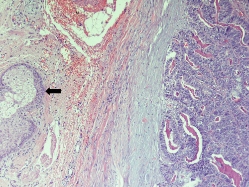
Histological section from skin specimen. Representative area of the skin lesion showing infiltration by moderately differentiated adenocarcinoma and a sebaceous gland (black arrow) (haematoxylin-eosin ×200).
Official websites use .gov
A
.gov website belongs to an official
government organization in the United States.
Secure .gov websites use HTTPS
A lock (
) or https:// means you've safely
connected to the .gov website. Share sensitive
information only on official, secure websites.
Histological section from skin specimen. Representative area of the skin lesion showing infiltration by moderately differentiated adenocarcinoma and a sebaceous gland (black arrow) (haematoxylin-eosin ×200).