
Figure 2.
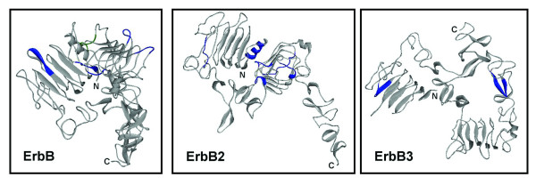
Localization of the identified UPMs in ErbB, ErbB2 and ErB3. The identified UPMs are labeled in blue on the crystal structures of extracellular domains in ErbB (PDB: 1NQL, 1210 residues), ErbB2 (PDB: 1N8Z, 1255 residues) and ErbB3 (PDB: 1M6B, 1342 residues). The EGF binding site on ErbB is labeled in green (S350-I356).