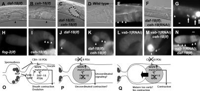
Figure 1.
daf-18 interacts with ceh-18 to regulate oocyte maturation. (A–C) Ovulation of the first oocyte in indicated strains. These animals were starved as L1s. The oocyte entered the spermatheca (arrowheads) in daf-18(lf) and ceh-18(lf) mutants, but failed to complete ovulation in daf-18(lf); ceh-18(lf) (dotted line). (D–G) Nomarski (D,F) and DAPI staining (E,G) images showing the Emo phenotype (arrows) of oocyte nuclei. Arrowheads indicate normal diakinesis nuclei. (H–N) MAP kinase activation in oocytes of unmated females of indicated strains. These strains all contain fog-2(lf). Stained oocytes are indicated with arrowheads in I, J, K, M, and N. (O–Q) Models to explain how daf-18 and ceh-18 pathways converge on MAP kinase to affect ovulation. (O) VAB-1 and CEH-18 constitute a mechanism that activates MAP kinase in response to sperm. Maturing oocytes send a signal to the sheath to stimulate contraction for ovulation. DAF-18 plays a cell-autonomous role in this mechanism by inhibiting MAP kinase in the oocyte through the PIP3:PIP2 ratio or AKT kinases, but not DAF-16. (P) When daf-18 and ceh-18 are inactivated, MAP kinase activity is deregulated in an additive manner. This may lead to uncoordinated signaling by the oocyte or to uncoordinated response by the sheath to the intense signal if animals are starved as L1s. (Q) Alternatively, the oocyte may mature too early in this condition for correct signaling. Bar, 0.01 mm (bar in G applies to F,G; bar in N applies to the rest).

