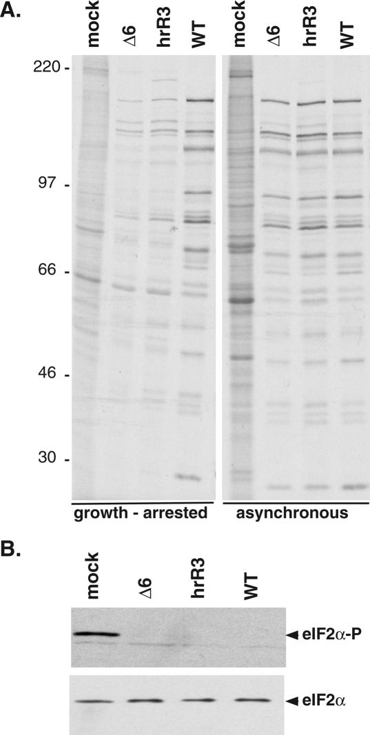
Figure 7.
Reduction in translation rates in quiescent cells infected with ICP6 mutants. (A) Growth-arrested or asynchronous, growing cells were infected (MOI = 10) with the indicated viruses. At 9 h post-infection, cells were radio-labeled for 1 h with 35S-methionine and cysteine. Total protein was subsequently isolated and fractionated by SDS-PAGE. The mobility of molecular weight standards (in kilodaltons) is shown to the left of the panel. (B) eIF2α phosphorylation in cells infected with ICP6 mutant viruses. Growth-arrested cells were infected with the indicated viruses as in A. After 10 h, total protein was isolated, fractionated by SDS-PAGE, and analyzed by immunoblotting with antibodies specific for either total eIF2α or phosphorylated eIF2α (eIF2α-P).

