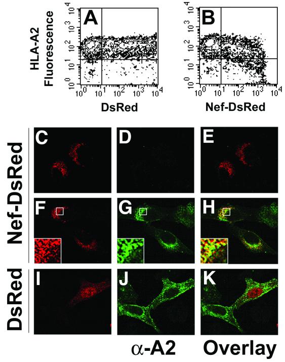
FIG. 1.
HIV-1 Nef-DsRed fusion protein causes reduction of cell surface MHC-I antigens, and colocalizes with MHC-I in the perinuclear area. (A and B) Nef-DsRed fusion protein downmodulates MHC-I. HeLa cells stably expressing HLA-A2 (HeLa-A2) were transiently transfected with plasmids encoding either DsRed (A) or a Nef-DsRed fusion protein (B). At 48 h posttransfection, HLA-A2 cell surface expression and DsRed protein expression were analyzed simultaneously by two-color flow cytometry. Crosshairs denote maximum fluorescence of negative controls for HLA-A2 staining (HeLa parental cells) and DsRed protein expression (mock-transfected HeLa-A2 cells). Results shown are representative of three independent experiments. (C through K) Nef-DsRed and MHC-I colocalize in the perinuclear region of cells. HeLa cells (C through E) or HeLa-A2 cells (F through K) were grown on glass slides and transiently transfected with plasmids encoding DsRed (I through K) or Nef-DsRed (C through H). At 24 h posttransfection, DsRed protein was detected by direct immunofluorescence (C, F, and I), and HLA-A2 was detected by indirect immunofluorescence using the HLA-A2-specific monoclonal antibody BB7.2 (D, G, and J). Enlargements of boxed regions are displayed as insets in the lower left corners of panels F through H. Images were collected using a Zeiss LSM 510 confocal laser scanning microscope, and single Z sections are displayed. Overlays and insets of DsRed and HLA-A2 images were generated using Adobe Photoshop imaging software. Results shown are representative of three independent experiments.

