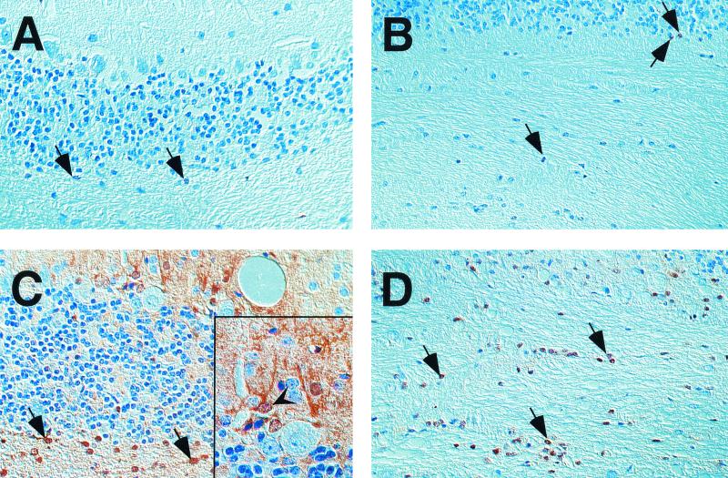
FIG. 4.
IHC for STAT1 revealed few immunoreactive cells in the cerebellar cortex (A) and white matter (B) of infected wild-type mice. In the respective brain regions of GF-IL-12 mice, strong expression of STAT1 was found (in the cortex [C] and the white matter [D]). Nuclear localization of STAT1 immunoreactivity in the cerebellar white matter (arrows). In infected GF-IL-12 mice there is additional immunoreactivity in the nuclei and the cytoplasm of cells resembling basket neurons (arrowhead). Magnification, ×40; magnification of inset, ×100.

