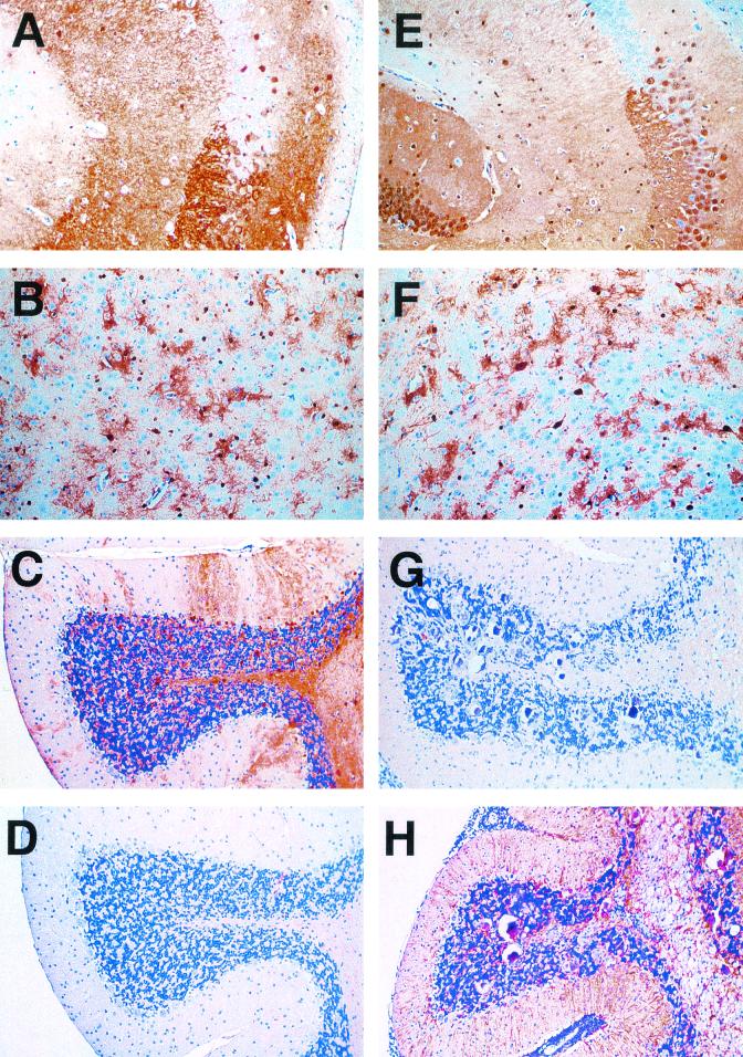
FIG. 5.
Reduced viral antigen levels in the cerebellum of GF-IL-12 mice. IHC staining for BDVp40 revealed virtually identical patterns in hippocampus (A and E) and thalamus (B and F) specimens from wild-type (A to D) and GF-IL-12 (E to H) mice. In contrast, in the cerebellum of GF-IL-12 mice (G), BDVp40 immunoreactivity was strongly reduced compared with that observed for wild-type mice (C). An inverse staining pattern was observed for GFAP. Immunoreactivity for GFAP was stronger in the cerebellum of GF-IL-12 mice (H) than in that of wild-type mice (D).

