
Figure 1.
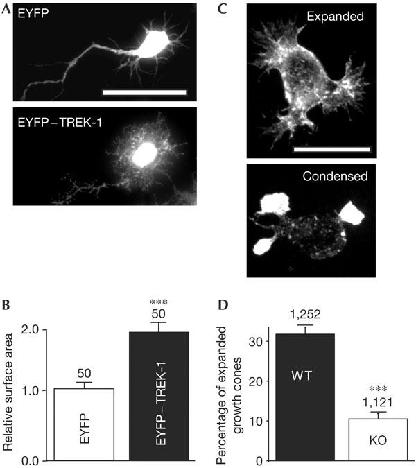
Expression of TREK-1 conditions the morphology of cultured neurons. (A) Young stage 2 hippocampal neurons were infected with Semliki forest virus bearing either EYFP (enhanced yellow fluorescent protein; top panel) or EYFP–TREK-1 (bottom panel). Scale bar, 20 μm. (B) A total of 50 individual neurons infected with each virus were randomly selected in a blind way and measured for surface area using the software ImageJ. The difference is significant (P<0.001). (C) Morphology of cultured embryonic striatal neurons from wild-type (WT) mice (top panel showing expanded growth cones) and TREK-1 knockout (KO) mice (bottom panel showing condensed growth cones). Neurons were stained with phalloidin. Scale bar, 10 μm. (D) Percentage of expanded growth cones in cultured neurons from TREK-1 WT (black column) and KO (white column) mice. Numbers of growth cones examined from four independent neuronal cultures (supplementary Fig 2 online) are indicated. The difference is significant (P<0.001).