
Figure 1.
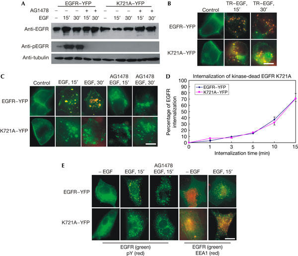
Internalization of kinase-dead epidermal growth factor receptor K721A. 293T cells (A–D) or Chinese hamster ovary (CHO) cells (E) were transiently transfected with epidermal growth factor receptor (EGFR)–YFP (yellow fluorescent protein) or K721A–YFP. (A) Immunoblotting. After EGF (100 ng/ml) stimulation, 293T cells were lysed and immunoblotted with indicated antibodies. (B) Intrinsic fluorescence. The cells were stimulated with Texas red (TR)-conjugated EGF (TR–EGF; 100 ng/ml), and the internalization of both EGFR and EGF was viewed by intrinsic fluorescence. (C) Indirect immunofluorescence. After EGF stimulation, the localization of EGFR (green) and phospho-tyrosine (pTyr; red) was shown by indirect immunofluorescence. (D) Quantitative analysis of EGFR–YFP and K721A–YFP internalization by flow cytometry. Data are the mean of at least three experiments performed in triplicate. (E) Indirect immunofluorescence analysis of the internalization of EGFR–YFP and K721A–YFP in CHO cells. CHO cells were transiently transfected with EGFR–YFP or K721A–YFP. After EGF (100 ng/ml) stimulation, the localization of EGFR (green) and pTyr (red) or EEA1 (red) was shown by indirect immunofluorescence. Scale bars, 20 μm.